44 compound microscope diagram without labels
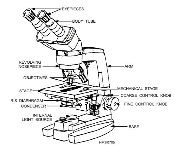
Label Microscope Diagram - EnchantedLearning.com Using the terms listed below, label the microscope diagram. arm - this attaches the eyepiece and body tube to the base. base - this supports the microscope. body tube - the tube that supports the eyepiece. coarse focus adjustment - a knob that makes large adjustments to the focus. diaphragm - an adjustable opening under the stage, allowing ... Labelled Diagram of Compound Microscope - Biology Discussion The below mentioned article provides a labelled diagram of compound microscope. Part # 1. The Stand: The stand is made up of a heavy foot which carries a curved inclinable limb or arm bearing the body tube. The foot is generally horse shoe-shaped structure (Fig. 2) which rests on table top or any other surface on which the microscope in kept.
PDF Parts of a Microscope Printables - Homeschool Creations typical student microscope -other microscopes will vary) •Which part of the microscope rotates so another person can look through the eyepiece without needing to move the microscope ? the head •What is the magnification level on the eyepiece of a microscope?10x (see objective lens magnification to see how these work together)

Compound microscope diagram without labels
Parts of a Compound Microscope and Their Functions Compound microscope magnification is determined by multiplying the eyepiece and objective powers. When viewed through a 5X eyepiece with a 10X objective, an item is magnified 5 x 10=50 times. The magnification is 10 x 45 = 450 times when using a 10X eyepiece and a 45X objective. How to Use the Compound Microscope Compound microscope - their parts and function - Microscopy4kids Compound microscopes have more than one lens to generate high magnification images of flat, thin specimens. 2. Eyepiece (10x) and Objective lenses (4x, 10x, 40x, 100x) are two major optical parts of a microscope. 3. Total magnification power is calculated by multiplying the magnification of the eyepiece and objective lens. 4. Microscope Parts and Functions Microscope Parts and Functions With Labeled Diagram and Functions How does a Compound Microscope Work?. Before exploring microscope parts and functions, you should probably understand that the compound light microscope is more complicated than just a microscope with more than one lens.. First, the purpose of a microscope is to magnify a small object or to magnify the fine details of a larger ...
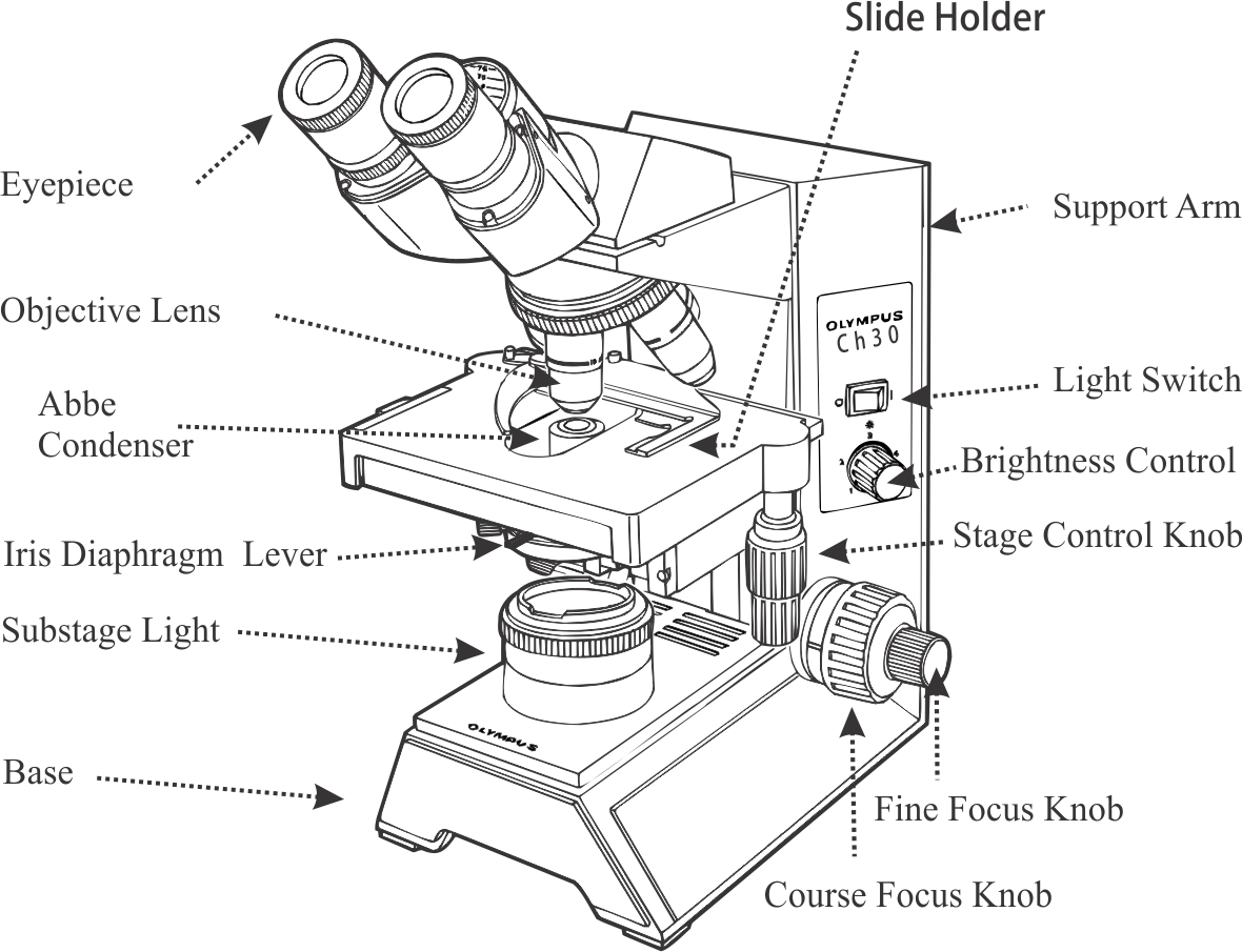
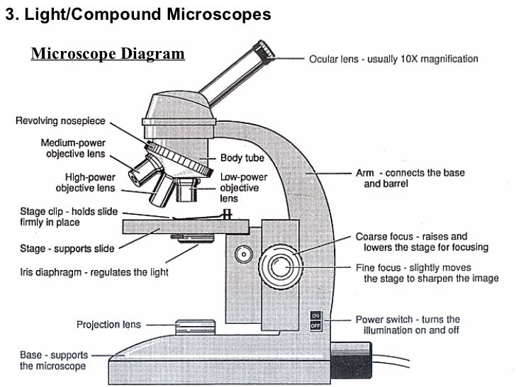
Compound microscope diagram without labels. Microscope, Microscope Parts, Labeled Diagram, and Functions The Iris Diaphragm is located above the condenser lens and below the microscope stage. The different sized holes in the diaphragm helps to vary the size of the cone and intensity of light that is projected upward into the slide. However, there is no set rule regarding which setting to use for a particular power. Working Principle and Parts of a Compound Microscope (with Diagrams) It holds the stage, body tube, fine adjustment and coarse adjustment. 5. Body Tube: It is usually a vertical tube holding the eyepiece at the top and the revolving nosepiece with the objectives at the bottom. The length of the draw tube is called 'mechanical tube length' and is usually 140-180 mm (mostly 160 mm). 6. Compound Light Microscope Diagram Worksheet - Google Groups Modern compound light microscopes under optimal conditions can we an average from 1000X to 2000X times the specimens original diameter Diagram. Label the parts of the microscope using the word... Labeling the Parts of the Microscope | Microscope World Resources Labeling the Parts of the Microscope This activity has been designed for use in homes and schools. Each microscope layout (both blank and the version with answers) are available as PDF downloads. You can view a more in-depth review of each part of the microscope here. Download the Label the Parts of the Microscope PDF printable version here.
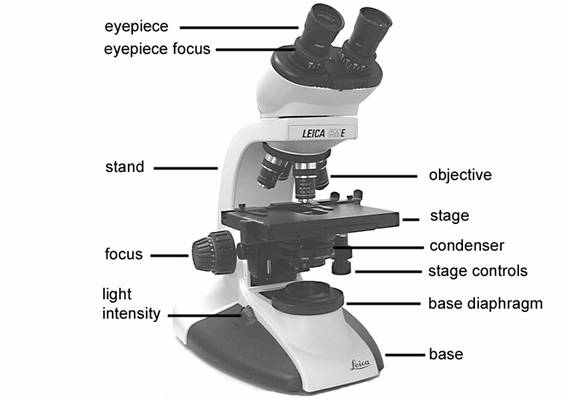
Label the microscope - Science Learning Hub In this interactive, you can label the different parts of a microscope. Use this with the Microscope parts activity to help students identify and label the main parts of a microscope and then describe their functions. Drag and drop the text labels onto the microscope diagram. Compound Microscope Parts Made Easy List and Diagram of Compound Microscope Parts: Head - The head is the uppermost part of the microscope that contains the eyepiece, tube, objective lens, and nosepiece. So all the optical parts of a compound microscope are in the head. Eyepiece - The eyepiece is the lens at the top, and the part you look through when using the microscope. Parts of a microscope with functions and labeled diagram Figure: Diagram of parts of a microscope There are three structural parts of the microscope i.e. head, base, and arm. Head - This is also known as the body. It carries the optical parts in the upper part of the microscope. Base - It acts as microscopes support. It also carries microscopic illuminators. Label A Microscope Teaching Resources | Teachers Pay Teachers This is a set of 3 tiered readings. Students will read a passage about the how to use a compound light microscope. Students will use textual evidence to answer questions and label the different parts of the microscope. It also allows students to gain prior knowledge about the compound microscope. Version A provides the most support for students.
Solved Label the image of a compound light microscope using - Chegg Step-by-step answer. Who are the experts? Experts are tested by Chegg as specialists in their subject area. We review their content and use your feedback to keep the quality high. Transcribed image text: Label the image of a compound light microscope using the terms provided. Compound Microscope- Definition, Labeled Diagram, Principle, Parts, Uses Alternatively, the magnification of the compound microscope is given by: m = D/ fo * L/fe where, D = Least distance of distinct vision (25 cm) L = Length of the microscope tube fo = Focal length of the objective lens fe = Focal length of the eye-piece lens Parts of a Compound Microscope Eyepiece And Body Tube. Compound Microscope Parts, Functions, and Labeled Diagram Compound Microscope Definitions for Labels. Eyepiece (ocular lens) with or without Pointer: The part that is looked through at the top of the compound microscope. Eyepieces typically have a magnification between 5x & 30x. Monocular or Binocular Head: Structural support that holds & connects the eyepieces to the objective lenses. A Study of the Microscope and its Functions With a Labeled Diagram To better understand the structure and function of a microscope, we need to take a look at the labeled microscope diagrams of the compound and electron microscope. These diagrams clearly explain the functioning of the microscopes along with their respective parts. Man's curiosity has led to great inventions. The microscope is one of them.
The Microscope - General Revision for GCSE
Diagram of a Compound Microscope - Biology Discussion A bright-field or compound microscope is primarily used to enlarge or magnify the image of the object that is being viewed, which can not otherwise be seen by the naked eye. Magnification may be defined as the degree of enlargement of the image of an object provided by the microscope.
Mycorrhizal Associations: Teaching Resources
Compound Microscope: Definition, Diagram, Parts, Uses, Working ... - BYJUS A compound microscope is defined as A microscope with a high resolution and uses two sets of lenses providing a 2-dimensional image of the sample. The term compound refers to the usage of more than one lens in the microscope. Also, the compound microscope is one of the types of optical microscopes.

Simple Microscope Labeled Diagram - Micropedia
16 Parts of a Compound Microscope: Diagrams and Video Once you have an understanding of the parts of the microscope it will be much easier to navigate around and begin observing your specimen, which is the fun part! The 16 core parts of a compound microscope are: Head (Body) Arm Base Eyepiece Eyepiece tube Objective lenses Revolving Nosepiece (Turret) Rack stop Coarse adjustment knobs
Compound Microscope Diagram With Labels - Micropedia
Compound Microscope Parts - Labeled Diagram and their Functions - Rs ... The term "compound" refers to the microscope having more than one lens. Basically, compound microscopes generate magnified images through an aligned pair of the objective lens and the ocular lens. In contrast, "simple microscopes" have only one convex lens and function more like glass magnifiers.
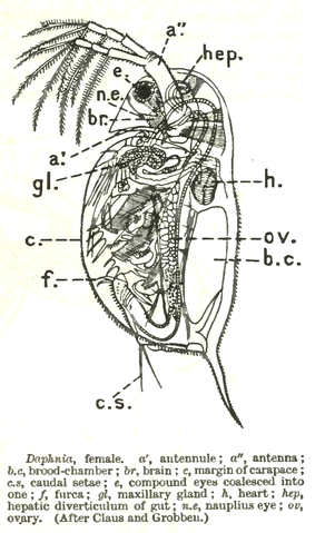
Water flea - Experiments on Microscopes 4 Schools
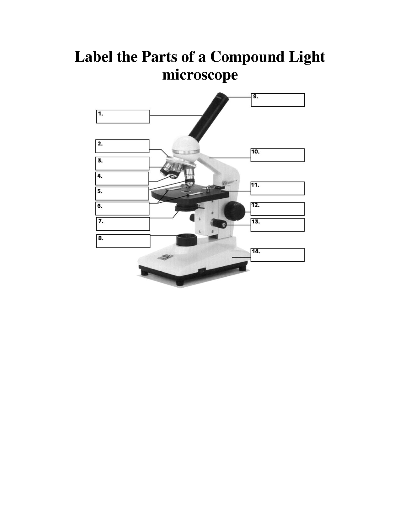
PDF Label compound microscope worksheet [clearBoth] [clearBoth] Microscope diagram without label After you've studied all the pieces of the composite microscope, it's time to put your brain to the test. Print an unmarked microscope chart and check that you can fill out all the blanks. [clearBoth] [clearBoth] Blank microscope diagram Next we have an empty microscope diagram.
[Solved] 1. Label the following microscope using the components ...
What is a Compound Microscope? - New York Microscope Company A compound microscope is an instrument that is used to view magnified images of small specimens on a glass slide. It can achieve higher levels of magnification than stereo or other low power microscopes and reduce chromatic aberration. It achieves this through the use of two or more lenses in the objective and the eyepiece.
The Cell
Compound Microscope - Types, Parts, Diagram, Functions and Uses A compound microscope captures an inverted image of the specimen because every time the light passes through the lens, the image's direction is flipped. The image always ends up inverted from the original. So, if you move the sample to the left, it moves in the right direction. Image 18: A comparison image between a simple and compound microscope.
8 Best Images of Lens Diagram Worksheet - Microscope with Labeled Parts ...
Compound Microscope: Parts of Compound Microscope - BYJUS The parts of the compound microscope can be categorized into: Mechanical parts; Optical parts (A) Mechanical Parts of a Compound Microscope. 1. Foot or base. It is a U-shaped structure and supports the entire weight of the compound microscope. 2. Pillar. It is a vertical projection. This stands by resting on the base and supports the stage. 3. Arm
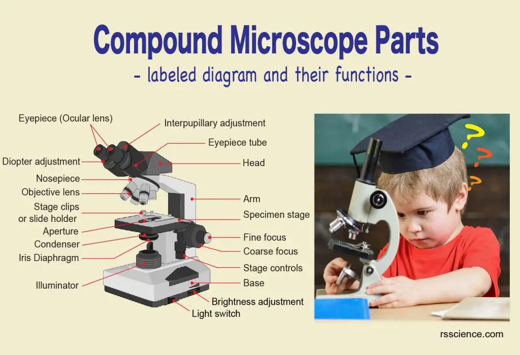
Compound Microscope Parts – Labeled Diagram and their Functions - Rs ...
Microscope Labeling - The Biology Corner Students label the parts of the microscope in this photo of a basic laboratory light microscope. Can be used for practice or as a quiz. ... The type of microscope used in most science classes is the _____ microscope. 18. You should carry the microscope by the _____ and the _____. 19. The objectives are attached to what part of the microscope ...
8 Best Images of Lens Diagram Worksheet - Microscope with Labeled Parts ...
Binocular Microscope Anatomy - Parts and Functions with a Labeled Diagram Now, I will discuss the details anatomy of the light compound microscope with the labeled diagram. Why it is called binocular: because it has two ocular lenses or an eyepiece on the head that attaches to the objective lens, this ocular lens magnifies the image produced by the objective lens. Binocular microscope parts and functions

The Microscope: Create a Labelled Diagram by ineedtoteachthat ...
Parts of the Microscope with Labeling (also Free Printouts) 5. Knobs (fine and coarse) By adjusting the knob, you can adjust the focus of the microscope. The majority of the microscope models today have the knobs mounted on the same part of the device. Image 5: The circled parts of the microscope are the fine and coarse adjustment knobs. Picture Source: bp.blogspot.com.

Microscope With Labels Clip Art at Clker.com - vector clip art online ...
Microscope Parts and Functions Microscope Parts and Functions With Labeled Diagram and Functions How does a Compound Microscope Work?. Before exploring microscope parts and functions, you should probably understand that the compound light microscope is more complicated than just a microscope with more than one lens.. First, the purpose of a microscope is to magnify a small object or to magnify the fine details of a larger ...

Label diagram of compound microscope - Science - The Fundamental Unit ...
Compound microscope - their parts and function - Microscopy4kids Compound microscopes have more than one lens to generate high magnification images of flat, thin specimens. 2. Eyepiece (10x) and Objective lenses (4x, 10x, 40x, 100x) are two major optical parts of a microscope. 3. Total magnification power is calculated by multiplying the magnification of the eyepiece and objective lens. 4.
Topic 3 - Microscopes
Parts of a Compound Microscope and Their Functions Compound microscope magnification is determined by multiplying the eyepiece and objective powers. When viewed through a 5X eyepiece with a 10X objective, an item is magnified 5 x 10=50 times. The magnification is 10 x 45 = 450 times when using a 10X eyepiece and a 45X objective. How to Use the Compound Microscope

Diagrams of sections of the Compound Microscopes at Science and Society ...

compound microscope 8th standard biology - YouTube

10 Best Images of Microscope Drawing Worksheet - Microscope Parts ...
Post a Comment for "44 compound microscope diagram without labels"